Myocardial Perfusion Scan, Resting
(Resting Thallium Scan, Cardiac Nuclear Imaging, Cardiolite® Scan, Sestamibi Scan)
What is a resting myocardial perfusion scan?
A myocardial perfusion scan is a type of nuclear medicine procedure. This means that a tiny amount of a radioactive substance, called a radionuclide (radiopharmaceutical or radioactive tracer), is used during the procedure to assist in the examination of the tissue under study. Specifically, the myocardial perfusion scan evaluates the heart’s function and blood flow.
A radionuclide is a radioactive substance used as a "tracer," which means it travels through the blood stream and is taken up (absorbed) by the healthy heart muscle tissue. On the scan, the areas where the radionuclide has been absorbed will show up differently than the areas that do not absorb it (due to decreased blood flow to the area or possible damage to the tissue from decreased or blocked blood flow).
A resting myocardial perfusion scan is used to assess the blood flow to the heart muscle (myocardium) and to determine what areas of the myocardium have decreased blood flow. This is done by injecting a radionuclide (thallium or technetium) into a vein in the arm or hand.
There are different types of radionuclides. When one type of radionuclide is used, areas of the myocardium that have blocked or partially blocked arteries will be seen on the scan as "cold spots," or "defects," because these areas will be unable to take in the radionuclide into the myocardium. Another type of radionuclide binds to the calcium that is released when a heart attack occurs, so it will accumulate in area(s) of injured heart tissue as a “hot spot” on the scan.
Other related procedures that may be used to diagnose heart disorders include resting and exercise electrocardiogram (ECG or EKG), Holter monitor, signal-averaged ECG, cardiac catheterization, chest x-ray, magnetic resonance imaging (MRI) of the heart, myocardial perfusion scan (stress), computed tomography (CT scan) of the chest, echocardiography, electrophysiological studies, radionuclide angiography, and ultrafast CT scan. Please see these procedures for additional information.
 Click Image to Enlarge
Coronary artery disease:
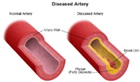
Coronary artery disease (CAD) is the narrowing of the coronary arteries (the blood vessels that supply oxygen and nutrients to the heart muscle), caused by a buildup of fatty material within the walls of the arteries. This buildup causes the inside of the arteries to become rough and narrowed, limiting the supply of oxygen-rich blood to the heart muscle.
To better understand how coronary artery disease affects the heart, a review of basic heart anatomy and function follows.
The heart is basically a pump. The heart is made up of specialized muscle tissue, called the myocardium. The heart's primary function is to pump blood throughout the body, so that the body's tissues can receive oxygen and nutrients and have waste substances taken away.
Like any pump, the heart requires fuel in order to work. The myocardium requires oxygen and nutrients, just like any other tissue in the body. However, the blood that passes through the heart's chambers is only passing through on its trip through the body - this blood does not give oxygen and nutrients to the myocardium. The myocardium receives its oxygen and nutrients from the coronary arteries, which lie on the outside of the heart.
 Click Image to Enlarge
When the heart tissue does not receive an adequate blood supply, it cannot function as well as it should. If the myocardium's blood supply is decreased for a length of time, a condition called ischemia may develop. Ischemia can decrease the heart's pumping ability, because the heart muscle is weakened due to a lack of food and oxygen.
Fortunately, the technology is available to restore blood flow to heart tissue when coronary artery blockages are diagnosed. One of several diagnostic procedures used to diagnose and evaluate coronary artery disease is the resting myocardial perfusion scan.
Possible indications for a resting myocardial perfusion scan may include, but are not limited to, the following:
- chest pain, either new onset or occurring over a period of days or longer
- following a heart attack (myocardial infarction, or MI)
- to assess blood flow to areas of the myocardium that have been reperfused (coronary artery blood flow restored) by bypass surgery, angioplasty (the opening of a coronary artery using a balloon or other method), or stent (a tiny expandable metal coil placed inside the artery to keep the artery open
The injection of the radionuclide may cause some slight discomfort. Allergic reactions to the radionuclide are rare.
If you are pregnant or suspect that you may be pregnant, you should notify your physician due to the risk of injury to the fetus from myocardial perfusion scan. If you are lactating, breastfeeding, you should notify your physician due to the risk of contaminating breast milk with radionuclide. Radiation exposure during pregnancy may lead to birth defects.
Patients who are allergic to or sensitive to medications, contrast dye, iodine, shellfish, tape, or latex should notify their physician.
There may be other risks depending upon your specific medical condition. Be sure to discuss any concerns with your physician prior to the procedure.
Certain factors or conditions may interfere with or affect the results of the test. These include, but are not limited to, the following:
- caffeine within 24 hours of the procedure
- digitalis, quinidine, or nitrate medications
- Your physician will explain the procedure to you and offer you the opportunity to ask any questions that you might have about the procedure.
- You will be asked to sign a consent form that gives your permission to do the test. Read the form carefully and ask questions if something is not clear.
- Notify your physician if you are allergic to or sensitive to medications, local anesthesia, contrast dyes, iodine, shellfish, tape, or latex.
- Fasting may be required before the procedure. Your physician will give you instructions as to how long you should withhold food and/or liquids. You should refrain from eating or drinking anything that contains caffeine for at least 24 hours prior to the procedure. Some prescription and over-the-counter medications contain caffeine and should be avoided. Some over-the-counter medications that contain caffeine include Anacin®, Excedrin®, and NoDoz®.
- Notify your physician of all medications (prescription and over-the-counter) and herbal supplements that you are taking.
- If you are pregnant or suspect that you may be pregnant, you should notify your physician.
- Notify your physician if you have a pacemaker.
- Based upon your medical condition, your physician may request other specific preparation.
A resting myocardial perfusion scan may be performed on an outpatient basis or as part of your stay in a hospital. Procedures may vary depending on your condition and your physician’s practices.
Generally, a resting myocardial perfusion scan follows this process:
- You will be asked to remove any jewelry or other objects that may interfere with the procedure.
- You will be asked to remove clothing and will be given a gown to wear.
- An intravenous (IV) line will be started in your hand or arm.
- You will be connected to an ECG machine with leads and a blood pressure cuff will be placed on your arm.
- You will lie flat on a table in the procedure room.
- The radionuclide will be injected into a vein in your arm or hand.
- After the medication has circulated through your body (10 to 60 minutes depending upon the radioactive tracer being used), the scanner will begin to take pictures of your heart. In a special kind of imaging test, called SPECT (single photon emission computed tomography), the scanner will rotate around you as it takes pictures.
- You will be lying flat on a table while the images of your heart are obtained. Your arms will be positioned on a pillow above your head. It will be necessary for you to lie very still while the images are being taken, as movement can adversely affect the quality of the images.
- If you experience any symptoms such as dizziness, chest pain, extreme shortness of breath, or severe fatigue at any point during the procedure, let the physician or technologist know.
- After the scan is complete, the IV line will be discontinued, and you will be allowed to leave, unless your physician instructs you differently.
You should move slowly when getting up from the scanner table to avoid any dizziness or lightheadedness from lying flat for the length of the procedure.
You will be instructed to drink plenty of fluids and empty your bladder frequently for 24 to 48 hours after the test to help flush the remaining radionuclide from your body.
The IV site will be checked for any signs of redness or swelling. If you notice any pain, redness, and/or swelling at the IV site after you return home following your procedure, you should notify your physician as this may indicate an infection or other type of reaction.
Your physician may give your additional or alternate instructions after the procedure, depending on your particular situation.
|


