Holter Monitor
(Continuous Electrocardiogram, Continuous ECG, Ambulatory ECG Monitoring)
What is a Holter monitor?
The Holter monitor is a type of electrocardiogram (ECG or EKG) used to monitor the ECG tracing continuously for a period of 24 hours or longer. An ECG is one of the simplest and fastest procedures used to evaluate the heart. Electrodes (small, plastic patches) are placed at certain locations on the chest, arms, and legs. When the electrodes are connected to an ECG machine by lead wires, the electrical activity of the heart is measured, interpreted, and printed out for the physician's information and further interpretation.
When symptoms such as dizziness, fainting, low blood pressure, prolonged fatigue, and palpitations continue to occur without a definitive diagnosis obtained with a resting ECG, an exercise ECG, or a signal-averaged ECG, your physician may request an ECG tracing to be run over a long period of time, using the Holter monitor.
Certain dysrhythmias/arrhythmias (abnormal heart rhythms), which can cause the symptoms noted above, may occur only intermittently, or may occur only under certain conditions, such as stress. Dysrhythmias of this type are difficult to obtain on an ECG tracing that only runs for a few minutes. Thus, the physician will request a Holter monitor to allow a better opportunity to capture any abnormal beats or rhythms that may be causing the symptoms. The Holter monitor records continuously for the entire period of 24 to 48 hours. Some Holter monitors may record continuously but also have an event monitor feature that you activate when symptoms begin to occur.
You will receive instructions regarding how long you will need to wear the recorder (usually 24 to 48 hours), how to keep a diary of your activities and symptoms during the test, and personal care/activity instructions.
What is an event monitor?
Event monitoring is very similar to Holter monitoring, and is often ordered for the same reasons. With an event monitor, you wear ECG electrode patches on your chest, and the electrodes are connected by wire leads to a recording device. Unlike the Holter monitor, however, which records continuously throughout the testing period of 24 to 48 hours, the event monitor does not record until you feel symptoms and trigger the monitor to record your ECG tracing at that time.
When you feel one or more symptoms, such as chest pain, dizziness, or palpitations, you push a button on the event monitor recorder. Some monitors have a feature (memory loop recorder) which captures a short period of time prior to the moment you triggered the recording and afterwards. This feature can help your physician determine more details about the possible change in your ECG at the time the symptoms started, and what was happening with your ECG just before you triggered the recorder. Other monitors, called "post-event recorders," simply start recording your ECG from the moment you trigger it.
Post-event recorders are quite small - some may even be worn on the wrist (similar to a wristwatch). Memory-loop recorders are about the size of a pager.
After you experience symptoms and record them, you will send the recording of the event to your physician or to a central monitoring center. This transmission is done over the telephone. You will be instructed regarding how to do this on the recorder. You will also keep a diary of your symptoms and corresponding activities (as done during the Holter monitoring procedure).
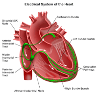
The heart's electrical conduction system:
The heart is, in the simplest terms, a pump made up of muscle tissue. Like all pumps, the heart requires a source of energy in order to function. The heart's pumping action comes from an intrinsic electrical conduction system.
 Click Image to Enlarge
An electrical stimulus is generated by the sinus node (also called the sinoatrial node, or SA node), which is a small mass of specialized tissue located in the right atrium (right upper chamber) of the heart.
The sinus node generates an electrical stimulus regularly at 60 to 100 times per minute under normal conditions. This electrical stimulus travels down through the conduction pathways (similar to the way electricity flows through power lines from the power plant to your house) and causes the heart's lower chambers to contract and pump out blood. The right and left atria (the two upper chambers of the heart) are stimulated first and contract a short period of time before the right and left ventricles (the two lower chambers of the heart).
The electrical impulse travels from the sinus node to the atrioventricular (AV) node, where impulses are slowed down for a very short period, then continues down the conduction pathway via the “bundle of His” into the ventricles. The “bundle of His” divides into right and left pathways to provide electrical stimulation to both ventricles.
This electrical activity of the heart is measured by an electrocardiogram. By placing electrodes at specific locations on the body (chest, arms, and legs), a graphic representation, or tracing, of the electrical activity can be obtained. Changes in an ECG from the normal tracing may indicate one or more of several heart-related conditions.
Some reasons for your physician to request a Holter monitor recording or event monitor recording include, but are not limited to, the following:
- to evaluate chest pain not reproduced with exercise testing
- to evaluate other signs and symptoms which may be heart-related, such as fatigue, shortness of breath, dizziness, or fainting
- to identify irregular heartbeats or palpitations
- to assess risk for future heart-related events in certain conditions, such as idiopathic hypertrophic cardiomyopathy (enlarged heart due to unknown reasons), post-heart attack with dysfunction of the left side of the heart, or Wolff-Parkinson-White syndrome (a condition in which an additional electrical pathway carries an impulse from the atria to the ventricles, causing rhythm problems)
- to assess the function of an implanted pacemaker
- to determine the effectiveness of therapy for complex arrhythmias
There may be other reasons for your physician to recommend the use of a Holter monitor.
The Holter monitor is a noninvasive method of assessing the heart’s function. Risks associated with the Holter monitor are rare.
Prolonged application of the adhesive electrode patches may cause tissue breakdown or skin irritation at the application site.
There may be other risks depending upon your specific medical condition. Be sure to discuss any concerns with your physician prior to wearing the monitor.
Certain factors or conditions may interfere with or affect the results of the Holter monitor reading. These include, but are not limited to, the following:
- close proximity to magnets, metal detectors, high-voltage electrical wires, and electrical appliances such as shavers, toothbrushes, and hair dryers
- smoking, certain medications
- excessive perspiration, which may cause the leads to loosen or detach
- Your physician will explain the procedure to you and offer you the opportunity to ask any questions that you might have about the reading.
- Fasting is not required.
- The area(s) where the electrodes are to be placed may be shaved.
- Based upon your medical condition, your physician may request other specific preparation.
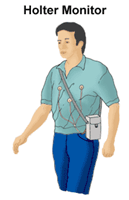
 Click Image to Enlarge
A Holter monitor recording is generally performed on an outpatient basis. Procedures may vary depending on your condition and your physician’s practices.
Generally, a Holter monitor recording follows this process:
- You will be asked to remove any jewelry or other objects that may interfere with the reading.
- You will be asked to remove clothing from the waist up in order to attach the electrodes to your chest. The technician will ensure your privacy by covering you with a sheet or gown and exposing only the necessary skin.
- If your chest, arms, or legs are very hairy, the technician may shave small patches of hair, as needed, so that the electrodes will stick closely to the skin.
- Electrodes will be attached to your chest, arms, and legs and the Holter monitor will be attached to the electrodes with lead wires. The monitor box may be worn over the shoulder like a shoulder bag, or it may clip to a belt or pocket.
- Once you have been hooked up to the monitor and given instructions, you can return to your usual activities, such as work, household chores, and exercise, unless your physician instructs you differently. This will allow your physician to identify problems that may only occur with certain activities.
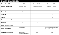
- You will be instructed to keep a diary of your activities during the recording period. You should write down the date and time of your activities, particularly if any symptoms, such as dizziness, palpitations, chest pain, or other previously-experienced symptoms, occur.
You should be able to resume your normal diet and activities, unless your physician instructs you differently.
Generally, there is no special care following a Holter monitor recording.
 Click Image to Enlarge
Notify your physician if you develop any signs or symptoms you had prior to the recording (e.g., chest pain, shortness of breath, dizziness, or fainting).
Your physician may give you additional or alternate instructions after the procedure, depending on your particular situation.
|


