Arrhythmias
An arrhythmia (also called dysrhythmia) is an abnormal rhythm of the heart, which can cause the heart to pump less effectively.
Arrhythmias can cause problems with contractions of the heart chambers by:
- not allowing the chambers to fill with an adequate amount of blood, because an electrical signal is causing the heart to pump too fast.
- not allowing a sufficient amount of blood to be pumped out to the body, because an electrical signal is causing the heart to pump too slowly or too irregularly.
In any of these situations, the heart may not be able to pump an adequate amount of blood to the body with each beat due to the arrhythmia's effects on the heart rate. The effects on the body are often the same, whether the heartbeat is too fast, too slow, or too irregular.
The following are the most common symptoms of arrhythmia. However, each child may experience symptoms differently. Symptoms may include:
- weakness
- fatigue
- palpitations
- low blood pressure
- dizziness
- fainting
The symptoms of arrhythmias may resemble other medical conditions or heart problems. Always consult your child's physician for a diagnosis.
Another indication of an arrhythmia is a change in the electrocardiogram (EKG or ECG) pattern. However, EKG changes are not seen unless an EKG test is performed or a child is being monitored in the hospital or other facility. Because symptoms such as those listed above may indicate the presence of an arrhythmia, an EKG is commonly done on children with one or more of the symptoms.
 Click Image to Enlarge
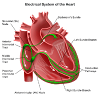
The heart is, in the simplest terms, a pump made up of muscle tissue. Like all pumps, the heart requires a source of energy in order to function. The heart's pumping action comes from an intrinsic electrical conduction system.
An electrical stimulus is generated by the sinus node (also called the sinoatrial node, or SA node), which is a small mass of specialized tissue located in the right atrium (right upper chamber) of the heart. The sinus node generates an electrical stimulus periodically (60-190 times per minute, depending on the age of the child and his/her activity level). This electrical stimulus travels down through the conduction pathways (similar to the way electricity flows through power lines from the power plant to your house) and causes the heart's lower chambers to contract and pump out blood. The right and left atria (the two upper chambers of the heart) are stimulated first and contract a short period of time before the right and left ventricles (the two lower chambers of the heart).
The electrical impulse travels from the sinus node to the atrioventricular (AV) node, where impulses are slowed down for a very short period, then continues down the conduction pathway via the bundle of His into the ventricles. The bundle of His divides into right and left pathways to provide electrical stimulation to both ventricles.
Normally, as the electrical impulse moves through the heart, the heart contracts about 60 to 100 times a minute. Each contraction of the ventricles represents one heartbeat. The atria contract a fraction of a second before the ventricles so their blood empties into the ventricles before the ventricles contract.
Under some conditions, almost all heart tissue is capable of starting a heartbeat, or becoming the "pacemaker," just like the sinus node. An arrhythmia may occur when:
- the heart's natural pacemaker (the sinus node) develops an abnormal rate or rhythm.
- the normal conduction pathway is interrupted.
- another part of the heart takes over as pacemaker.
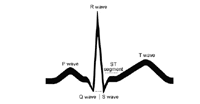
The electrical activity of the heart is measured by an electrocardiogram (ECG or EKG). By placing electrodes at specific locations on the body (chest, arms, and legs), a graphic representation, or tracing, of the electrical activity can be obtained. Changes in an ECG from the normal tracing can indicate arrhythmias, as well as other heart-related conditions.
Almost everyone knows what a basic ECG tracing looks like. But what does it mean?
 Click Image to Enlarge
- The first little upward notch of the ECG tracing is called the "P wave." The P wave indicates that the atria (the two upper chambers of the heart) are electrically stimulated to pump blood to the ventricles.
- The next part of the tracing is a short downward section connected to a tall upward section. This next part is called the "QRS complex." This part indicates that the ventricles (the two lower chambers of the heart) are electrically stimulated to pump out blood to the body.
- The next short flat segment is called the "ST segment." The ST segment indicates the amount of time from the end of the contraction of the ventricles to the beginning of the "T wave".
- The next upward curve is called the "T wave." The T wave indicates the recovery period of the ventricles.
When your child's physician studies your child's ECG, he/she looks at the size and length of each part of the EKG. Variations in size and length of the different parts of the tracing may be significant.
The tracing for each lead of a 12-lead ECG will look different, but will have the same basic components as described above. Each lead of the 12-lead ECG is "looking" at a specific part of the heart from different angles. Variations in a lead may indicate a problem with the part of the heart associated with that particular lead.
An atrial arrhythmia is an arrhythmia caused by abnormal function of the sinus node, or by the development of another atrial pacemaker within the heart tissue that takes over the function of the sinus node.
A ventricular arrhythmia is an arrhythmia caused by abnormal function of the sinus node, an interruption in the electrical conduction pathways, or the development of another area within the heart tissue that takes over the function of the sinus node.
Arrhythmias can also be classified as slow (bradyarrhythmia) or fast (tachyarrhythmia). "Brady-" means slow, while "tachy-" means fast.
Listed below are some of the more common arrhythmias:
The symptoms of various arrhythmias may resemble other medical conditions or heart problems. Always consult your child's physician for a diagnosis.
In addition to a complete medical history and physical examination of your child, there are several different types of procedures that may be used to diagnose arrhythmias. Some of these procedures include the following:
Specific treatment for arrhythmias will be determined by your child's physician based on:
- your child's age, overall health, and medical history
- extent of the condition
- your child' s tolerance for specific medications, procedures, or therapies
- expectations for the course of the condition
- your opinion or preference
Arrhythmias may be present but cause few, if any, problems. In this case, your child's physician may elect not to treat the arrhythmia. However, when the arrhythmia causes symptoms, there are several different options for treatment. Your child's physician will choose an arrhythmia treatment based on the type of arrhythmia, the severity of symptoms being experienced, and the presence of other conditions (i.e., diabetes, kidney failure, heart failure) which can affect the course of the treatment.
Treatments may include:
- lifestyle modifications
Factors such as stress, caffeine, or alcohol can cause arrhythmias. Your child's physician may order the elimination of caffeine, alcohol (teens and young adults), or any other substance believed to be causing the problem. If stress is suspected as a cause, your child's physician may recommend stress-reduction measures such as an exercise program or family therapy.
- medication
There are various types of medications which may be used to treat arrhythmias. If your child's physician chooses to use medication, the decision of which medication to use will be determined by the type of arrhythmia, other conditions which may be present, and other medications already being used by your child.
- cardioversion
In this procedure, a small, electrical shock is delivered to the heart through the chest to stop certain, very fast, arrhythmias such as atrial fibrillation, supraventricular tachycardia, or sinus tachycardia. Your child is given medication to help him/her relax, and is then connected to an EKG monitor which is also connected to the cardioversion device. The small, electrical shock is delivered at a precise point during the EKG cycle.
- ablation
This is an invasive procedure done in the electrophysiology laboratory, and involves a small, thin tube (catheter) being inserted into the heart through a vessel in the groin or arm. The procedure is done in a manner similar to the electrophysiology studies (EPS) described above. Once the site of the arrhythmia has been determined by EPS, the catheter is moved to the site. By use of a technique such as radiofrequency ablation (very high frequency radio waves are applied to the site, heating the tissue until the site is destroyed) or cryoablation (an ultra-cold substance is applied to the site, freezing the tissue and destroying the site), the site of the arrhythmia may be destroyed.
- pacemaker
A permanent pacemaker is a small device that is implanted under the skin and sends electrical signals to start or regulate a slow heartbeat. A permanent pacemaker may be used to make the heart beat if the heart's natural pacemaker (the sinoatrial, or SA, node) is not functioning properly and has developed an abnormal heart rate or rhythm or if the electrical pathways are blocked. Pacemakers are typically used for slow arrhythmias such as sinus bradycardia, sick sinus syndrome, or heart block.
In infants and young children, pacemakers are usually placed in the abdomen. The wires that connect the pacemaker to the heart are placed on the outside surface of the heart. This position is beneficial because the fat in the abdomen protects the pacemaker and pacemaker wires from injury that might occur during everyday childhood activities such as climbing and falling.
School-aged children and adolescents may have the pacemaker placed in the shoulder area just under the collarbone. The pacemaker wires are often placed inside the superior vena cava, a large vein that connects to the right atrium, and then guided inside the heart.
- implantable cardioverter defibrillator
An implantable converter defibrillator (ICD) is a small device, similar to a pacemaker, that is implanted under the skin, often in the shoulder area just under the collarbone. An ICD senses the rate of the heartbeat. When the heart rate exceeds a rate programmed into the device, it delivers a small, electrical shock to the heart to slow the heart rate. Many newer ICDs can also function as a pacemaker by delivering an electrical signal to regulate a heart rate that is too slow. ICDs are typically used for fast arrhythmias such as ventricular tachycardia.
- surgery
Surgical treatment for arrhythmias is usually done only when all other appropriate options have failed. Surgical ablation is a major surgical procedure requiring general anesthesia. The chest is opened, exposing the heart. The site of the arrhythmia is located, then destroyed or removed in order to eliminate the arrhythmia.
Click here to view the
Online Resources of Heart Center
|


