Vascular Studies
(Carotid, Arm, and Leg Arterial and Venous Studies, Carotid Ultrasound, Venous Doppler Studies, Arterial Doppler Studies, Pulse Volume Recordings, PVRS)
What are vascular studies?
Vascular studies are a noninvasive (the skin is not pierced) procedure used to assess the blood flow in arteries and veins. A transducer (like a microphone) sends out ultrasonic sound waves at a frequency too high to be heard. When the transducer is placed on the skin at certain locations and angles, the ultrasonic sound waves move through the skin and other body tissues to the blood vessels, where the waves echo off of the blood cells. The transducer picks up the reflected waves and sends them to an amplifier, which makes the ultrasonic sound waves audible.
Vascular studies can utilize one of these special types of ultrasound technology, as listed below:
- Doppler ultrasound
This Doppler technique is used to measure and assess the flow of blood through the blood vessels. The amount of blood pumped with each beat is an indication of the size of a vessel’s opening. Also, Doppler can detect abnormal blood flow within a vessel, which can indicate a blockage caused by a blood clot, a plaque, or inflammation.
- color Doppler
Color Doppler is an enhanced form of Doppler ultrasound technology. With color Doppler, different colors are used to designate the direction of blood flow. This simplifies the interpretation of the Doppler technique.
 Click Image to Enlarge
To assess blood flow in the limbs, pulse volume recordings (PVRs) may be performed. Blood pressure cuffs are inflated on the limb and blood pressure in the limb is measured using the Doppler transducer.
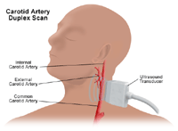
To assess the carotid arteries in the neck, a carotid duplex scan may be performed. This type of Doppler examination provides a 2-dimensional (2D) image of the arteries so that the structure of the arteries and location of an occlusion can be determined, as well as the degree of blood flow.
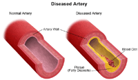
A carotid artery duplex scan is a type of vascular ultrasound study done to assess occlusion (blockage) or stenosis (narrowing) of the carotid arteries of the neck and/or the branches of the carotid artery. Plaque (a build up of fatty materials), a thrombus (blood clot), and other substances in the blood stream may cause a disturbance in the blood flow through the carotid arteries.
Other related procedures that may be used to assess the heart and circulatory system include resting and exercise electrocardiogram (ECG or EKG), Holter monitor, signal-averaged ECG, cardiac catheterization, chest x-ray, computed tomography (CT scan) of the chest, electrophysiological studies, magnetic resonance imaging (MRI) of the heart, myocardial perfusion scans, radionuclide angiography, and ultrafast CT scan. Please see these procedures for additional information.
Vascular conditions:
The arteries bring oxygen and other nutrients to the cells of the body. The veins take away the blood after the cells have taken in the oxygen and nutrients and given up their waste products, such as carbon dioxide. If blood flow is decreased to any part of the body, that area does not get enough oxygen and nutrients and is unable to get rid of its waste products adequately.
 Click Image to Enlarge
Decreased blood flow can occur in the arteries and veins anywhere in the body, such as the neck and brain. When the neck arteries (carotid arteries) become occluded, symptoms such as dizziness, confusion, drowsiness, headache, and/or a brief loss of ability to speak or move, may be the early warning signs of a possible stroke (brain attack). More severe symptoms, such as sudden sharp headache, loss of vision in one eye, sudden loss of ability to move arms, legs, or one side of the body, sudden forceful vomiting, or sudden decreased level of consciousness may mean that a stroke is imminent.
Some conditions which may affect blood flow include, but are not limited to, the following:
- atherosclerosis - a gradual clogging of the arteries over many years by fatty materials and other substances in the blood stream
- aneurysm - a dilation of a part of the heart muscle or the aorta (the large artery that carries oxygenated blood out of the heart to the rest of the body), which may cause weakness of the tissue at the site of the aneurysm
- embolus or thrombus - clots in blood vessels may be either an embolus (a small mass of material such as fat globules, air, clusters of bacteria, or even foreign matter such as a piece of metal from a bullet) or a thrombus (a blood clot)
- inflammatory conditions - an inflammation within a blood vessel may occur as a result of trauma (physical trauma, such as from a fall, or chemical trauma, such as from an irritating medication being introduced into the vessel), infection, or an autoimmune disorder (e.g., polyarteritis, Raynaud's disease, and aortic arch syndrome)
- varicose veins - occur when the veins of the circulatory system in the legs are exposed over time to pressure that causes stress on the walls and valves of the veins
Any of these conditions may cause decreased blood flow in arteries and/or veins. Some symptoms that may occur when blood flow decreases to the legs include, but are not limited to, the following:
- leg pain and/or weakness during exertion (known as claudication)
- swelling
- soreness, tenderness, redness, and/or warmth in the leg(s)
- pale and cool skin; may even be grayish or blue
- numbness or tingling
- rest pain (pain in the foot that occurs when sitting or lying down and is relieved by standing)
If the physician suspects that a person may have decreased blood flow somewhere in the peripheral (arms, legs, and/or neck) circulation, vascular studies may be performed.
Reasons for which vascular studies may be performed include, but are not limited to, the following:
- evaluation of signs and symptoms which may suggest decreased blood flow in the arteries and/or veins of the neck, legs, or arms
- evaluation of previous procedures that were performed to restore blood flow to an area
- evaluation of a vascular dialysis device, such as an A-V fistula in the arm
There may be other reasons for your physician to recommend a vascular study.
There is no radiation used and generally no discomfort from the application of the ultrasound transducer to the skin.
For some patients, having to lie still on the examination table for the length of the procedure may cause some discomfort or pain.
There may be other risks depending upon your specific medical condition. Be sure to discuss any concerns with your physician prior to the procedure.
Certain factors or conditions may interfere with a vascular study. These factors include, but are not limited to, the following:
- smoking for at least an hour before the test, as smoking causes blood vessels to constrict
- severe obesity
- cardiac dysrhythmias/arrhythmias (irregular heart rhythms)
- cardiac disease
- Your physician will explain the procedure to you and offer you the opportunity to ask any questions that you might have about the procedure.
- Generally, no prior preparation, such as fasting or sedation, is required.
- Your physician may give you specific instructions about smoking and consuming caffeine. You may be asked to refrain from smoking for at least two hours before the test, as smoking causes blood vessels to constrict. You may also be asked to refrain from consuming caffeine in any form for about two hours prior to the test.
- Based upon your medical condition, your physician may request other specific preparation.
A vascular study may be performed on an outpatient basis or as part of your stay in a hospital. Procedures may vary depending on your condition and your physician's practices.
Generally, a vascular study follows this process:
- You will be asked to remove any jewelry or other objects that may interfere with the procedure. You may wear your glasses, dentures, or hearing aid if you use any of these.
- If you are asked to remove clothing, you will be given a gown to wear.
- You will lie on an exam table or bed.
- A clear gel will be placed on the skin at locations where the pulse is expected to be heard.
- The Doppler transducer will be pressed against the skin and moved around over the area of the artery or vein being studied.
- When blood flow is detected, you will hear a "whoosh, whoosh" sound. The probe will be moved around to compare blood flow in different areas of the artery or vein.
- For arterial studies of the legs, blood pressure cuffs will be applied in three positions on the leg in order to compare the blood pressure between different areas of the leg. The cuff around the thigh will be inflated first, and the blood pressure will be determined with the Doppler transducer placed just below the cuff.
- The cuff around the calf will be inflated, and the blood pressure will be determined as with the thigh cuff.
- The cuff around the ankle will be inflated, and the blood pressure will be determined as in steps #6 and #7.
- The blood pressure will be taken in the arm on the same side as the leg that was just studied and used to determine the degree of any occlusion of arterial flow in the legs.
- Once the procedure has been completed, the gel will be removed from the skin.
The technologist will use all possible comfort measures and complete the procedure as quickly as possible to minimize any discomfort.
You may resume your usual diet and activities unless your physician advises you differently.
Generally, there is no special type of care following a vascular study. However, your physician may give you additional or alternate instructions after the procedure, depending on your particular situation.
|