Exercise Electrocardiogram
(Exercise ECG, Exercise EKG, Stress Test)
What is an exercise electrocardiogram?
An electrocardiogram (ECG or EKG) is one of the simplest and fastest procedures used to evaluate the heart. Electrodes (small, plastic patches) are placed at certain locations on the chest, arms, and legs. When the electrodes are connected to an ECG machine by lead wires, the electrical activity of the heart is measured, interpreted, and printed out for the physician's information and further interpretation.
An exercise ECG is performed to assess the heart's response to stress or exercise. The ECG is monitored while a person is exercising on a treadmill or stationary bike. While this procedure is seldom used for young children, it may be very useful in evaluating adolescents and young adults.
An ECG tracing will be taken at certain points during the test in order to compare the effects of increasing stress on the heart. Periodically, the incline and treadmill speed will be increased in order to make exercise more difficult for the person being tested. If the person is riding a bicycle, he/she will pedal faster against increased resistance. In either circumstance, the person will exercise until reaching a target heart rate (determined by the physician based on age and physical status) or until unable to continue due to fatigue, shortness of breath, chest pain, or other symptoms.
Other related procedures that may be used to assess the heart include resting electrocardiogram (ECG), Holter monitor, signal-averaged ECG, cardiac catheterization, chest x-ray, computed tomography (CT scan) of the chest, echocardiography, electrophysiological studies, magnetic resonance imaging (MRI) of the heart, myocardial perfusion scans, radionuclide angiography, and ultrafast CT scan. Please see these procedures for additional information.
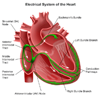
The heart's electrical conduction system:
The heart is, in the simplest terms, a pump made up of muscle tissue. Like all pumps, the heart requires a source of energy in order to function. The heart's pumping action comes from an intrinsic electrical conduction system.
 Click Image to Enlarge
An electrical stimulus is generated by the sinus node (also called the sinoatrial node, or SA node), which is a small mass of specialized tissue located in the right atrium (right upper chamber) of the heart.
The sinus node generates an electrical stimulus regularly at 60 to 100 times per minute under normal conditions. This electrical stimulus travels down through the conduction pathways (similar to the way electricity flows through power lines from the power plant to your house) and causes the heart's lower chambers to contract and pump out blood. The right and left atria (the two upper chambers of the heart) are stimulated first and contract a short period of time before the right and left ventricles (the two lower chambers of the heart).
The electrical impulse travels from the sinus node to the atrioventricular (AV) node, where impulses are slowed down for a very short period, then continues down the conduction pathway via the “bundle of His” into the ventricles. The “bundle of His” divides into right and left pathways to provide electrical stimulation to both ventricles.
This electrical activity of the heart is measured by an electrocardiogram. By placing electrodes at specific locations on the body (chest, arms, and legs), a graphic representation, or tracing, of the electrical activity can be obtained. Changes in an EKG from the normal tracing may indicate one or more of several heart-related conditions.
Reasons for your physician to request an exercise ECG include, but are not limited to, the following:
- to determine limits for safe exercise in patients who are entering a cardiac rehabilitation program and/or those who are recovering from a cardiac event, such as a heart attack (myocardial infarction, or MI) or heart surgery
- to assess leg pain with exercise (also called intermittent claudication) in patients with suspected occlusion in the legs' circulatory system
- to evaluate blood pressure during exercise
- to assess stress or exercise tolerance in patients with known or suspected coronary artery disease
There may be other reasons for your physician to recommend an exercise ECG.
Because of the stress the heart incurs during the procedure, there is a small chance for chest pain, heart attack, high blood pressure, irregular heartbeats, dizziness, nausea, and extreme fatigue. Notify your physician if you have the following conditions:
- aneurysm - a dilation of a part of the heart muscle or the aorta (the large artery that carries oxygenated blood out of the heart to the rest of the body) which may cause a weakness of the tissue at the site of the aneurysm
- unstable angina (uncontrolled chest pain)
- severe heart valve disease
- severe congestive heart failure
- recent myocardial infarction (also called MI, or heart attack)
- severe hypertension (high blood pressure)
- uncontrolled irregular heartbeats
- pericarditis (an inflammation or infection of the sac which surrounds the heart)
- severe anemia (low red blood cell count)
If you are pregnant or suspect that you may be pregnant, you should notify your physician.
Prolonged application of the adhesive electrode patches may cause tissue breakdown or skin irritation at the application site.
There may be other risks depending upon your specific medical condition. Be sure to discuss any concerns with your physician prior to the procedure.
Certain factors or conditions may interfere with or affect the results of the test. These include, but are not limited to, the following:
- intake of a heavy meal, caffeine, and/or smoking prior to the procedure
- high blood pressure
- electrolyte abnormalities, such as too much or too little potassium, magnesium, and/or calcium in the blood
- certain medications
- heart valve disease
- enlarged left ventricle
- Your physician or the technician will explain the procedure to you and offer you the opportunity to ask any questions that you might have about the procedure.
- You will be asked to sign a consent form that gives your permission to do the procedure. Read the form carefully and ask questions if something is not clear.
- You will be asked to fast for a few hours before the procedure. You should not smoke for two hours prior to the procedure.
- If you are pregnant or suspect that you may be pregnant, you should notify your physician.
- Notify your physician of all medications (prescription and over-the-counter) and herbal supplements that you are taking.
- Wear flat shoes that are comfortable for walking and loose-fitting pants or shorts. Women should wear a short-sleeved top that fastens in the front for ease of attaching the ECG electrodes to the chest.
- The area(s) where the electrodes are to be placed may be shaved.
- Based upon your medical condition, your physician may request other specific preparation.
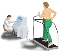
 Click Image to Enlarge
An exercise ECG may be performed on an outpatient basis or as part of your stay in a hospital. Procedures may vary depending on your condition and your physician’s practices.
Generally, an exercise ECG follows this process:
- You will be asked to remove any jewelry or other objects that may interfere with the procedure.
- You will be asked to open your blouse or shirt in the front (men may be asked to remove their shirts). The technician will ensure your privacy by covering you with a sheet or gown and exposing only the necessary skin.
- If your chest, arms, or legs are very hairy, the technician may shave small patches of hair, as needed, so that the electrodes will stick closely to the skin.
- Electrodes will be attached to your chest, arms, and legs.
- The lead wires will be attached to the skin electrodes.
- Once the leads are attached, the technician may key in identifying information about you into the machine's computer.
- A blood pressure cuff will be attached to your arm while you are sitting down. Initial, or baseline, ECG and blood pressure readings will be taken while you are sitting down and standing up.
- You will be instructed on how to walk on the treadmill. Alternately, you may exercise on a bicycle. You will be told to let the technician, physician, or nurse know if you begin to have any chest pain, dizziness, lightheadedness, extreme shortness of breath, nausea, headache, leg pains, or other symptoms during exercise.
- You will begin to exercise at a minimal level. The intensity of the exercise will be gradually increased on the treadmill by increasing the incline and speed of the treadmill every few minutes.
- ECG and blood pressure readings will be taken periodically during the exercise to measure how well your heart and body are responding to the exercise.
- The exercise will end once you have reached a target heart rate (determined by the physician based on your age and physical condition). The test may also be stopped if you develop severe symptoms such as chest pain, dizziness, nausea, severe shortness of breath, severe fatigue, or elevated blood pressure.
- Once you have reached your target heart rate, the rate of exercise will be slowed for a "cool down" period to help avoid any nausea or cramping from sudden stopping of exercise.
- You will sit in a chair and your ECG and blood pressure will be monitored until they return to normal or near-normal. This may take 10 to 20 minutes.
- Once your ECG and blood pressure readings are acceptable to the physician, the ECG electrodes and blood pressure cuff will be removed. You may then put on your shirt or blouse.
You should be able to resume your normal diet and activities, unless your physician instructs you differently.
Generally, there is no special care following an exercise ECG.
You may feel tired for several hours or longer after the procedure, particularly if you do not normally exercise. Otherwise, you should feel normal within a few hours after the procedure, if not sooner. If your fatigue lasts longer than a day, you should notify your physician.
Notify your physician if you develop any signs or symptoms you had prior to the test (e.g., chest pain, shortness of breath, dizziness, or fainting).
Your physician may give you additional or alternate instructions after the procedure, depending on your particular situation.
|