Chest X-Ray
(Chest Radiography, CXR)
What is a chest x-ray?
A chest x-ray is a type of diagnostic radiology procedure used to examine the chest and the organs and structures located in the chest. Chest x-rays may be used to assess the lungs, as well as the heart (either directly or indirectly) by looking at the heart itself. Certain conditions of the heart may cause changes in the lungs and/or the vessels of the lungs. Changes in the normal structure of the heart, lungs, and/or lung vessels may indicate disease or other conditions.
Chest x-rays may provide important information regarding the size, shape, contour, and anatomic location of the heart, lungs, bronchi, great vessels (aorta, aortic arch, pulmonary arteries), mediastinum (an area in the middle of the chest separating the lungs), and the bones (cervical and dorsal spine, clavicles, shoulder girdle, and ribs).
What are x-rays?
X-rays use invisible electromagnetic energy beams to produce images of internal tissues, bones, and organs on film. X-rays are made by using external radiation to produce images of the body, its organs and other internal structures for diagnostic purposes. X-rays pass through body tissues onto specially treated plates (similar to camera film) and a “negative” type picture is made (the more solid a structure is, the whiter it appears on the film).
Depending on the results of the chest x-ray, additional tests or procedures may be requested by your physician for further diagnostic information.
Other related procedures that may be used to diagnose problems of the chest and respiratory tract include chest fluoroscopy, chest ultrasound, computed tomography (CT scan) of the chest, lung biopsy, lung scans, mediastinoscopy, positron emission tomography (PET scan) of the chest, pleural biopsy, thoracentesis, sinus x-rays, pulmonary angiogram, bronchoscopy, and bronchography. Please see these procedures for additional information.
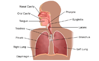
Anatomy of the respiratory system:
 Click Image to Enlarge
The respiratory system is made up of the organs involved in the interchanges of gases, and consists of the:
- nose
- pharynx
- larynx
- trachea
- bronchi
- lungs
The upper respiratory tract includes the:
- nose
- nasal cavity
- ethmoidal air cells
- frontal sinuses
- maxillary sinus
- larynx
- trachea
The lower respiratory tract includes the lungs, bronchi, and alveoli.
What are the functions of the lungs?
The lungs take in oxygen, which cells need to live and carry out their normal functions. The lungs also get rid of carbon dioxide, a waste product of the body's cells.
The lungs are a pair of cone-shaped organs made up of spongy, pinkish-gray tissue. They take up most of the space in the chest, or the thorax (the part of the body between the base of the neck and diaphragm).
The lungs are enveloped in a membrane called the pleura.
The lungs are separated from each other by the mediastinum, an area that contains the following:
- the heart and its large vessels
- trachea (windpipe)
- esophagus
- thymus
- lymph nodes
The right lung has three sections, called lobes. The left lung has two lobes. When you breathe, the air enters the body through the nose or the mouth. It then travels down the throat through the larynx (voice box) and trachea (windpipe) and goes into the lungs through tubes called main-stem bronchi.
One main-stem bronchus leads to the right lung and one to the left lung. In the lungs, the main-stem bronchi divide into smaller bronchi and then into even smaller tubes called bronchioles. Bronchioles end in tiny air sacs called alveoli.
Chest x-rays may be used to assess heart status (either directly or indirectly) by looking at the heart itself, as well as the lungs. Changes in the normal structure of the heart, lungs and/or lung vessels may indicate disease or other conditions.
Conditions that may be assessed with a chest x-ray include, but are not limited to, the following:
- heart enlargement (which can occur with congenital heart defects or cardiomyopathy)
- pericardial effusion - a build up of excess fluid in-between the heart and the membrane that surrounds it, often due to inflammation
- pleural effusion - a collection of blood or fluid around the lung
- pneumonia, persistent cough, and other lung conditions
- aneurysms - ballooning of the walls of the great blood vessels, such as the aorta
- bone fractures
- calcification of heart structures (such as heart valves or aorta)
- tumors or cancer
- herniation of the diaphragm (the breathing muscle, the diaphragm, moves out of place)
- pleuritis - inflammation of the lining of the lung
- pulmonary edema (“fluid in the lungs,” which can occur with congenital heart disease or congestive heart failure)
Other reasons for performing a chest x-ray may include:
- as part of the physical assessment before hospitalization and/or surgery or as part of a complete physical examination
- to assess symptoms of conditions related to the heart or lungs
- to assess progression of a condition and/or effectiveness of treatments
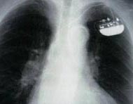
- to check the position of implanted pacemaker wires and other internal devices such as central venous catheters, endotracheal tubes, chest tubes, or nasogastric tubes
- to check status of lungs and chest cavity after surgery
There may be other reasons for your physician to recommend a chest x-ray.
The amount of radiation used during a chest x-ray procedure is considered minimal; therefore, the risk for radiation exposure is very low.
If you are pregnant or suspect that you may be pregnant, you should notify your physician. Radiation exposure during pregnancy may lead to birth defects.
There may be other risks depending upon your specific medical condition. Be sure to discuss any concerns with your physician prior to the procedure.
- The physician will explain the procedure to you and offer you the opportunity to ask any questions that you might have about the procedure.
- If you are pregnant or suspect that you may be pregnant, you should notify your physician.
- Generally, no prior preparation, such as fasting or sedation, is required.
- Dress in clothes that permit access to the area to be tested or that are easily removed.
- Notify the radiologic technologist if you have any body piercing on your chest.
- Based upon your medical condition, your physician may request other specific preparation.
A chest x-ray may be performed on an outpatient basis or as part of your stay in a hospital. Procedures may vary depending on your condition and your physician’s practices.
 Chest X-ray with Implanted Pacemaker
Generally, a chest x-ray follows this process:
- You will be asked to remove any clothing, jewelry, or other objects that may interfere with the procedure.
- You will be given a gown to wear.
- The particular view that the physician orders will determine how you are positioned for the x-ray such as lying, sitting, or standing. You will be positioned carefully so that the desired view of the chest is obtained. The physician will also specify the number of films to be made.
- For a standing or sitting film, you will stand or sit in front of the x-ray plate. You will be asked to roll your shoulders forward, take in a deep breath, and hold it until the x-ray exposure is made. For patients who are unable to hold their breath, the radiologic technologist will take the picture at the appropriate time by watching the breathing pattern.
- It will be important for you to remain still during the exposure, as any movement will blur the film.
- For a side-angle view of the chest, you will be asked to turn to your side and raise your arms above your head. You will be instructed to take in a deep breath and hold it as the x-ray exposure is made.
- The radiologic technologist will step behind a protective window while the images are being made.
While the x-ray procedure itself causes no pain, the manipulation of the body part being examined may cause some discomfort or pain, particularly in the case of a recent injury or invasive procedure such as surgery. The radiologic technologist will use all possible comfort measures and complete the procedure as quickly as possible to minimize any discomfort or pain.
Generally, there is no special type of care after a chest x-ray. However, your physician may give you additional or alternate instructions after the procedure, depending on your particular situation.
|