Overview of Pacemakers
An implanted pacemaker is a small device that is implanted under the skin and sends electrical signals to start or regulate a slow heartbeat. An implanted pacemaker may be used to stimulate the heartbeat if the heart's natural pacemaker (the sinoatrial, or SA, node) is not functioning properly, has developed an abnormally slow heart rate or rhythm, or if the electrical pathways are blocked.
Rhythm problems are common in teens and young adults with congenital heart disease due to changes in scar tissue and other results from prior surgeries and procedures.
Children's pacemakers may be placed under the skin in one of several locations. Young children (infants, toddler, preschool, and young school-aged children) often have the pacemaker generator placed in the abdomen, since the fatty tissue found there can help protect the generator from normal everyday childhood activities such as playing. As a child gets older (nearing adolescence), the generator is often placed in the shoulder area, just under the collarbone.
An implantable cardioverter defibrillator (ICD) is a small device, similar to a pacemaker, that is implanted under the skin, often in the shoulder area, just under the collarbone. An ICD senses the rate of the heartbeat. When the heart rate exceeds a rate programmed into the device, the ICD delivers a small, electrical shock to the heart to slow the heart rate. Many newer ICDs can also function as a pacemaker by delivering an electrical signal to regulate a heart rate that is too slow or pace out of a rapid rhythm.
When the heart's natural pacemaker has a dysfunction, the signals it sends out may become erratic: either too slow, too fast, or too irregular to stimulate adequate contractions of the heart chambers. When the heartbeat becomes erratic, it is referred to as an arrhythmia (an abnormal rhythm of the heart, which can cause the heart to pump less effectively).
ICDs may be recommended for persons with significant ventricular rhythm problems which could pose a risk for sudden death. These rhythm problems are often associated with worsening heart failure.
Arrhythmias can cause problems with contractions of the heart chambers by:
- not allowing the chambers to fill with an adequate amount of blood because the electrical signal is causing the heart to pump too fast.
- not allowing a sufficient amount of blood to be pumped out to the body because the electrical signal is causing the heart to pump too slow or too irregularly.
 Click Image to Enlarge
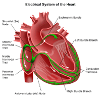
The heart is, in the simplest terms, a pump made up of muscle tissue. Like all pumps, the heart requires a source of energy in order to function. The heart's pumping action comes from an intrinsic electrical conduction system.
An electrical stimulus is generated by the sinus node (also called the sinoatrial node, or SA node), which is a small mass of specialized tissue located in the right atrium (right upper chamber) of the heart. The sinus node generates an electrical stimulus periodically (60-190 times per minute, depending on the age of the child and his/her activity level). This electrical stimulus travels down through the conduction pathways (similar to the way electricity flows through power lines from the power plant to your house) and causes the heart's lower chambers to contract and pump out blood. The right and left atria (the two upper chambers of the heart) are stimulated first and contract a short period of time before the right and left ventricles (the two lower chambers of the heart).
The electrical impulse travels from the sinus node to the atrioventricular (AV) node, where impulses are slowed down for a very short period, then continues down the conduction pathway via the bundle of His into the ventricles. The bundle of His divides into right and left pathways to provide electrical stimulation to both ventricles.
Normally, as the electrical impulse moves through the heart, the heart contracts about 60 to 100 times a minute. Each contraction of the ventricles represents one heartbeat. The atria contract a fraction of a second before the ventricles so their blood empties into the ventricles before the ventricles contract.
Under some conditions, almost all heart tissue is capable of starting a heartbeat, or becoming the "pacemaker," just like the sinus node. An arrhythmia may occur when:
- the heart's natural pacemaker (the sinus node) develops an abnormal rate or rhythm.
- the normal conduction pathway is interrupted.
- another part of the heart takes over as pacemaker.
In any of these situations, the body may not receive enough blood because the heart cannot pump out an adequate amount with each beat as a result of the arrhythmia's effects on the heart rate. The effects on the body are often the same, however, whether the heartbeat is too fast, too slow, or too irregular.
A permanent pacemaker has two components, including the following:
- a pulse generator which has a sealed lithium battery and an electronic circuitry package. The pulse generator produces the electrical signals that make the heart beat. Many pulse generators also have the capability to receive and respond to signals that are sent by the heart itself.
- one or two wires (also called leads). Leads are insulated, flexible wires that conduct electrical signals to the heart from the pulse generator. The leads may also relay signals from the heart to the pulse generator. One end of the lead is attached to the pulse generator and the electrode end of the lead is positioned in the atrium (the upper chamber of the heart) or in the ventricle (the lower chamber of the heart).
Older pacemakers sent out electrical signals at a constant rate, regardless of the heart's own rate. Pacemaker technology is now much more advanced. Today, pacemakers can "sense" when the heart's natural rate falls below the rate that has been programmed into the pacemaker's circuitry.
 Click Image to Enlarge
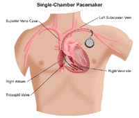
Pacemaker leads may be positioned in the atrium, ventricle, or both, depending on the condition requiring the pacemaker to be inserted.
- An atrial arrhythmia (an arrhythmia caused by a dysfunction of the sinus node or the development of another atrial pacemaker within the heart tissue that takes over the function of the sinus node) may be treated with an atrial permanent pacemaker whose lead wire is located in the atrium.
- A ventricular arrhythmia (an arrhythmia caused by a dysfunction of the sinus node, an interruption in the conduction pathways, or the development of another pacemaker within the heart tissue that takes over the function of the sinus node) may be treated with a ventricular pacemaker whose lead wire is located in the ventricle.
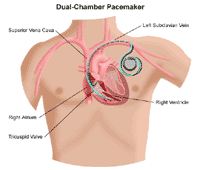
- It is possible to have both atrial and ventricular arrhythmias, and there are pacemakers which have lead wires positioned in both the atrium and the ventricle. There may be one lead wire for each chamber, or one lead wire may be capable of sensing and pacing both chambers.
 Click Image to Enlarge
- An ICD has a lead wire that is positioned in the ventricle, as it is used primarily for fast ventricular arrhythmias.
Pacemakers that pace either the right atrium or the right ventricle are called "single-chamber" pacemakers. Pacemakers that pace both the right atrium and right ventricle of the heart and require two pacing leads are called "dual-chamber" pacemakers.
Pacemaker/ICD insertion is done in the hospital, either as a short-stay surgical procedure, or in the cardiac catheterization or electrophysiology laboratory. The child is given medication to help him/her relax during the procedure.
In older children and teenagers who receive a transvenous pacemaker, a small incision is made just under the collarbone. The pacemaker/ICD lead(s) is inserted into the heart through a blood vessel which runs under the collarbone. This procedure is usually performed in the catheterization laboratory.
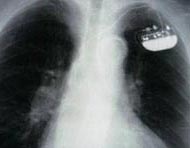
 The picture above is a chest x-ray. The
large, white space in the middle is the
heart. The dark spaces on either side are
the lungs. The small object in the upper
corner is an implanted pacemaker.In younger children, the pacemaker may be placed into the abdomen through a small incision. A second incision is made in the chest to visualize the heart. The lead(s) are guided to the heart, then placed on the heart's surface. This procedure is usually performed in the operating room. Once the procedure has been completed, the child goes through a recovery period of several hours and often is allowed to go home the day after the procedure.
After receiving a pacemaker or ICD, you will receive an identification card from the manufacturer that includes information about your child's specific model of pacemaker and the serial number. You should carry this card with you at all times so that the information is always available to any healthcare professional who may have reason to examine and/or treat your child. A medical identification bracelet or necklace can also be worn by your child to alert others about the pacemaker or ICD in case of emergency.
Click here to view the
Online Resources of Heart Center
|